| Catalog No. | HY546106 |
| Brand | abinScience |
| Tested applications | WB, FCM |
| Species reactivity | Human |
| Form | Liquid |
| Storage buffer | 0.01M PBS pH 7.4 |
| Purity | >95% purity as determined by SDS-PAGE. |
| Isotype | IgG1-kappa |
| Applications | ELISA, Bioactivity: FACS, Functional assay, Research in vivo |
| Target | Meconium antigen 100, CEACAM5, CEA, CD66e, Carcinoembryonic antigen-related cell adhesion molecule 5, Carcinoembryonic antigen |
| Purification | Protein A/G purified from cell culture supernatant. |
| Endotoxin level | < 10 EU/mg |
| Expression system | Mammalian cells |
| Accession | P06731 |
| Stability and Storage | Use a manual defrost freezer and avoid repeated freeze thaw cycles. Store at 2 to 8°C for frequent use. Store at -20 to -80°C for twelve months from the date of receipt. |
| Alternative name | SGM-101, SGM101, SGM101 |
| Clone ID | SGM 101 |
| Note | For research use only. Not suitable for clinical or therapeutic use. |
| Isotype Control | Human IgG1 Recombinant Isotype Control Antibody (13R4) (HV080507) |
Anti-Human CEACAM-5/CD66e Reference Antibody (SGM 101, RUO)
Overview
Images
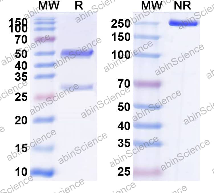 SDS-PAGE |
SDS-PAGE for Research Grade Anti-Human CD66e/CEA/CEACAM5 (SGM 101). |
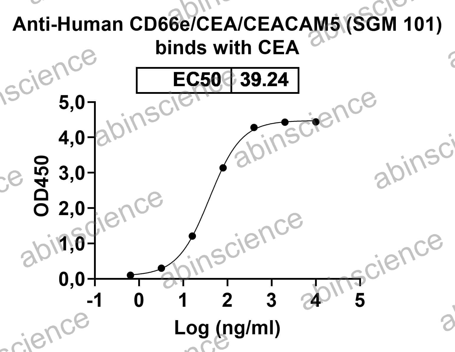 Bioactivity |
Detects recombinant human CD66e/CEA/CEACAM5 (Cat No.: HY546011) in indirect ELISA. |
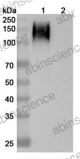 Western blot |
Western blot analysis was performed using anti-CD66e/CEA/CEACAM5 monoclonal antibody at 1ug/mL on various samples. Lane 1: recombinant human CD66e/CEA/CEACAM5 (Catalog No: HY546011) Lane 2: negative control |
 Flow cytometry |
Flow-cytometry using PE anti-human CD66e antibody. HPAC cells were stained with an irrelevant antibody (Blue Histogram) or an PE anti-human CD66e monoclonal antibody (Catalog HY546106, Yellow Histogram) at a concentration of 5 µg/ml for 30 mins at RT. After washing, and cells analysed on a NovoCyte Flow Cytometer. |
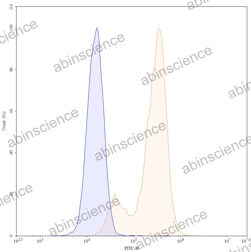 Flow cytometry |
Flow-cytometry using FITC anti-human CD66e antibody. HPAC cells were stained with an irrelevant antibody (Blue Histogram) or an FITC anti-human CD66e monoclonal antibody (Catalog HY546106, Yellow Histogram) at a concentration of 5 µg/ml for 30 mins at RT. After washing, and cells analysed on a NovoCyte Flow Cytometer. |